Lab: Investigating the action of 6 enzymes
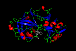
| Enzymes are proteins that catalyze (that is, increase or decrease the rates of) chemical reactions. In enzymatic reactions, the molecules at the beginning of the process are called substrates, and they are converted into different molecules, called the products. Almost all processes in a biological cell need enzymes to occur at significant rates. Since enzymes are selective for their substrates and speed up only a few reactions from among many possibilities, the set of enzymes made in a cell determines which metabolic pathways occur in that cell. This extract is licensed under the Creative Commons Attribution-ShareAlike license. It uses material from the article "Enzyme", retrieved 23 Feb 2011. |
This lab offers an opportunity to investigate the action of six different enzymes.
Contents
Prep
- Learn how enzymes work by reviewing the concepts presented at the following sites:
- The basics: Lab 2: Enzyme Catalysis (see Concepts 1-6)
- Very good simulation: Enzyme Action and the Hydrolysis of Sucrose
- Somewhat technical: Reactions and Enzymes
- Technical explanation: Enzymes
- Enzyme problem set that includes tutorials with each problem (some of the problems are easier than others): Energy, Enzymes and Catalysis Problem Set
- Printout the instructions below for the six activities included in this lab.
Amylase
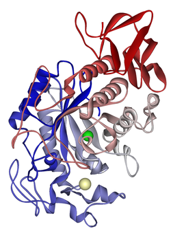
Amylase is an enzyme that breaks down starch into sugar. Amylase is present in human saliva, where it begins the chemical process of digestion. Foods that contain much starch but little sugar, such as rice and potato, taste slightly sweet as they are chewed because amylase turns some of their starch into sugar in the mouth. Plants and some bacteria also produce amylase. In seeds, amylase initiates the breakdown of starch to glucose during germination. [1]
The iodine test is used to test for the presence of starch. Iodine solution — iodine dissolved in an aqueous solution of potassium iodide — reacts with starch producing a purple black color. Elemental iodine solutions will stain starches due to iodine's interaction with the coil structure of the polysaccharide. As starch is broken down or hydrolyzed into smaller carbohydrate units, in this case by the enzyme amylase, the purple-black color is not produced. Therefore, this test can determine completion of hydrolysis when a color change does not occur.[2][3]
In this demonstration you will observe what happens when amylase from animals and plants is put into contact with starch molecules.[4]
Materials needed:
- Plain agar powder
- Clear dishes – petri dishes or glass dishes
- Q-tips
- Iodine, potassium iodide (IKI) solution* (or commercial tincture of iodine, diluted 1:4 with water)
- Untreated corn seeds
- Glucose test strips
- *Iodine, potassium iodide solution is harmful if inhaled or swallowed. It causes burns to skin and eyes. Goggles should be worn when using this solution. See links to Hands-on Science Project: Chemical Safety Database for further safety information:
Procedure:
- Prepare 4 starch-agar plates. Allow to cool.
- Using a glucose test strip, test one of the agar plates for the presence of sugar. Record results.
- Label the 4 starch-agar plates: "human", "soaked seeds", "boiled seeds", "dry seeds"
- Prepare corn seeds:
- 5 soaked
- 5 boiled
- 5 plain
- Add amylase to plates:
- For human amylase, put the end of a Q-tip in your mouth, then use that tip to write a message on a starch plate.
- Use a sharp razor blade to cut each corn grain longitudinally and place, cut surface down, onto the surface of the agar, spaced 2cm from each other. (You may wish to dissect out the embryo.)
- Allow plates to stand for 30 minutes.
- Remove corn and rinse plate gently with distilled water.
- Flood all 4 plates with IKI solution, swish around as color develops, rinse with distilled water.
- Using a glucose test strip, test one of the clear areas for the presence of sugar.
- Observe/record results for human saliva and for each of the corn seed conditions.
Data table
Before beginning the activity, design a data table to record the results for each element in the demonstration.
Papain
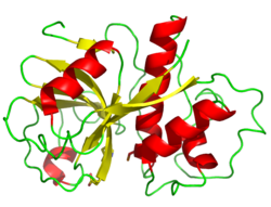
Papain is a cysteine protease enzyme present in papaya. Papain belongs to a family of enzymes which break down proteins, by preaking the peptide bonds between amino acids.Papain may be used to break down tough meat fibers and has been utilized for thousands of years in its native South America. It is sold as a component in powdered meat tenderizer available in most supermarkets. Papain, in the form of a meat tenderizer such as Adolph's, made into a paste with water, is also a home remedy treatment for jellyfish, bee, and wasps stings; mosquito bites; and possibly stingray wounds, breaking down the protein toxins in the venom.[5]
Gelatin is a protein produced by partial hydrolysis of collagen extracted from the boiled bones, connective tissues, organs and some intestines of animals such as domesticated cattle, pigs, and horses. Gelatin is a solid when cooled, and a liquid when heated.[6]
In this activity you will investigate the action of meat tenderizer, containing the enzyme papain, on gelatin and its ability to "gel."[4]
Materials needed:
- Powdered gelatin (pkg), e.g., Knox
- Beaker (150 ml) or other heat-resistant container
- Teaspoon (or balance, if weighing)
- Meat tenderizer, e.g., Adolph's
- Stirring rods (3)
- Test tubes (2)
- Ice water
- Hot plate or stove
- Distilled water (100 ml)
Procedure:
The beginning of the procedure is provided below:
- Prepare a gelatin solution by heating 1 teaspoon (3.0 g) of gelatin in 100 ml distilled water until dissolved. (Gently mix, do not boil.)
- Cool the gelatin solution to room temperature.
Using the prepared gelatin solution and the materials listed above, design an experiment to test the effect of meat tenderizer on the ability of the gelatin to "gel".
Data table
Before beginning design a data table for recording the results.
Catalase
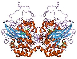
Catalase is a common enzyme found in nearly all living organisms that are exposed to oxygen, where it functions to catalyze the decomposition of hydrogen peroxide to water and oxygen.- 2 H2O2 → 2 H2O + O2
Catalase has one of the highest turnover numbers of all enzymes; one catalase enzyme can convert 40 million molecules of hydrogen peroxide to water and oxygen each second. (Hydrogen peroxide is a harmful by-product of many normal metabolic processes: to prevent damage, it must be quickly converted into other, less dangerous substances.)[7]
In this activity you will investigate the action of hydrogen peroxide when put in contact with living and non-living organisms. You may also wish to investigate the effect of temperature on the reaction.[4]
Materials needed:
- Test tubes or cups (one for each material to be tested)
- Hydrogen peroxide (H2O2) (3% solution)
- Distilled water (for control)
- Assorted living tissue: sliced banana, sliced raw potato, ground meat, liver, yeast cells, ground young leaves
- Assorted non-living material: piece of baked potato, cooked liver, etc.(Note that if using rocks or sand, some will “bubble.”)
- Water baths at various temperatures
Procedure:
Design an experiment from which you can deduce the action of catalase (present in living organisms) on hydrogen peroxide. If interested, include temperature, hot, room temperature, cold, as an independent variable. (Note that heat will degrade the hydrogen peroxide.)
Data table
Before beginning design a data table for recording the results.
Invertase
Invertase is an enzyme that catalyzes the hydrolysis (breakdown) of sucrose, a disaccharide, also known as table sugar, into two monosaccharides, fructose and glucose. For industrial use, invertase is usually obtained from yeast.[8]
In this activity you will investigate the action of invertase (from yeast) in the presence of sucrose.[4]
Materials needed:
- Yeast
- Sucrose (0.25 M solution)
- Distilled water (100 ml)
- Stirring rod
- Beaker
- Test tubes or cups
- Pipet or syringe to measure 3 ml
- Glucose test strips
- Balance or teaspoon
Procedure:
- Prepare yeast by mixing 1 teaspoon (3 g) dry yeast with 20 ml of distilled water. Let stand for 20 minutes.
- Test the sucrose solution with a glucose test strip for the presence of glucose.
- Fill each of two test tubes 1/3 full with sucrose solution.
- Label the two test tubes, one of which will contain the yeast mixture, with the other serving as the control.
- Add 3 ml yeast suspension to one tube. Mix.
- Add 3 ml distilled water to the other tube. Mix.
- After 10 minutes use a glucose test strip to test each test tube for the presence of glucose.
(For best results, do not go over the recommended incubation period.)
Data table
Before beginning design a data table for recording the results.
Chymosin
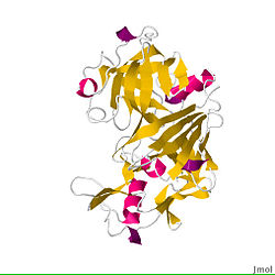
Chymosin (also termed rennin) is an enzyme produced by cows in the lining of the abomasum (the fourth and final, chamber of the stomach). Chymosin is the active ingredient in rennet, which is used in making cheese.[9] Placed in milk, chymosin breaks down a protein called kappa casein which keeps milk in liquid form. The breaking down of kappa casein leads to coagulation, or "curdling" of the milk, which becomes the basis for cheese.[10] Rennet is the active "thickening" agent in Junket, a milk-based dessert, available at many US grocery stores.In this activity you will investigate the action of chymosin, as a component of the enzyme complex rennet, on milk protein.[4]
Materials needed:
- Junket
- Milk (do not use UHT long shelf-life type)
- Distilled Water (50ml)
- Beaker (100 ml)
- Stirring rods (3)
- Test tubes or cups (2)
Procedure:
Dissolve 1 tablet of junket with 1 T water, creating a rennet solution.
Design an experiment to investigate the action of chymosin on milk. (Note that the action of chymosin on milk requires 10 min incubation at room temperature.)
Data table
Before beginning design a data table for recording the results.
Pepsin
Pepsin is an enzyme whose precursor form (pepsinogen) is released by the chief cells in the stomach and which degrades food proteins into peptides. Pepsin functions best in acidic environments, in particular environments with a pH of 1.5 to 2. Pepsin denatures if the pH is more than 5.0.[11]In this activity you will investigate the action of pepsin on the "coagulated" protein contained in hard-boiled egg white.[4]
Materials needed:
- Egg white (hard-boiled)
- Test tubes or cups (6)
- Test tube rack
- Dietary supplement containing pepsin
- Vinegar
- Sodium bicarbonate (baking soda)
- Distilled water
- Metric ruler
- Knife
Procedure:
- Label 6 test tubes or small cups: A to F
- Add the following solutions to each test tube:
- A: Pepsin tablet dissolved in 5mL distilled water
- B: Pepsin tablet dissolved in 5mL white vinegar (5% acid solution)
- C: 5mL white vinegar (5% acid solution)
- D: Pepsin tablet dissolved in 5mL 0.5% sodium bicarbonate solution
- E: 5 mL 0.5% sodium bicarbonate solution
- F: 5 mL distilled water
- Drop a small (2 mm) cube of egg white into each tube.
- Incubate at room temperature for approximately 12 hours. (The speed of this reaction can be increased by using very thin strips of egg white and/or incubating at 30°C.)
- Examine tubes for the presence of the egg white.
Data table
Before beginning design a data table for recording the results.
Some ideas for conclusion/discussion
- How are all of these experiments/demonstrations related?
- For each enzyme, how do the results of the experiment relate to the action of the enzyme in its natural environment?
- When you designed your own experiment did it turn out as you expected? Would you recommend any changes to your procedure?
- What limitations exist in these experiments? (Hint: does the experiment/demonstration provide proof of an enzyme's action?)
- What characteristics of the environment may affect the action of an enzyme? Did any of the experiments address one or more of these characteristics?
Thought questions
- The packaging for a gelatin dessert in the USA includes the note: "Do not use fresh or frozen pineapple, kiwi, gingerroot, papaya, figs or guava. Gelatin will not set." Any thoughts on what these foods have in common?
- The pH (a measure of the acidity or basicity of a solution) may affect the action of an enzyme. Which other enzymes might be good candidates for investigating the effect of pH on the enzyme's action? Why?
- Why would cooking a plant or piece of meat affect the activity of catalase, but not the activity of pepsin?
References
- ↑ Amylase. In Wikipedia, retrieved, 23 Feb 2011.
- ↑ Iodine test. In Wikipedia, retrieved 23 Feb 2010.
- ↑ Logul's iodine. In Wikipedia, retrieved 23 Feb 2010.
- ↑ 4.0 4.1 4.2 4.3 4.4 4.5 Aspects adapted from Miller, S. B. (1992). Simple enzyme experiments. In Tested studies for laboratory teaching, Volume 6 (C.A. Goldman, S.E. Andrews, P.L. Hauta, and R. Ketchum, Editors). Proceedings of the 6th Workshop/Conference of the Association for Biology Laboratory Education (ABLE).
- ↑ Papain. In Wikipedia, retrieved 24 Feb 2011.
- ↑ Gelatin. In Wikipedia, retrieved 24 Feb 2011.
- ↑ Catalase. In Wikipedia, retrieved, 25 Feb 2011.
- ↑ Invertase. In Wikipedia, retrieved, 25 Feb 2011.
- ↑ Chymosin. In Wikipedia, retrieved, 26 Feb 2011.
- ↑ cheeseforme. In isbibbio, retrieved 26 Feb 2011.
- ↑ Pepsin. In Wikipedia, retrieved, 25 Feb 2011.
